
The Autoimmune Disease Myth
Autoimmune disease affects 23.5 million people in the US and millions more worldwide, yet the medical industry has no idea what causes it. Mainstream theory states that the immune system has gone haywire and is attacking the patient’s own tissues. But of course this is yet another mythology of pop culture, and the real cause of the problem known as “autoimmune disease” is estrogen dominance – and it’s entirely reversible.
Despite this, doctors like to blame autoimmune disease on genetics even though not a single gene has been found to trigger any autoimmune disease.
If autoimmune disease is genetic then why does an injection of the hormone estrogen causes the same “autoantibodies” found in autoimmune patients to appear?
As with most diseases, the scientific evidence paints an entirely different picture than what the public is told. The evidence shows that the body’s own immune system isn’t attacking its own tissues at all, but rather cleaning up a mess caused by dead cellular debris. When talking about autoimmune disease, what we’re really talking about is widespread tissue damage in the body.
In this presentation, you’ll learn in complete detail what autoimmune disease is and how to prevent or reverse it safely and effectively with a number of approaches, including dietary modifications, natural supplements, medical drugs and red light therapy.
(Click here to watch on YouTube)

TABLE OF CONTENTS
Mainstream Theory of Autoimmune Disease
The mainstream theory of autoimmune disease is that for some unknown reason the immune system has begun attacking the body. A doctor might tell you researchers are still searching for the genes that cause autoimmune disease but the reality is they’re never going to find them, because what’s happening has nothing to do with genetics.
Examples of autoimmune diseases:
- Rheumatoid Arthritis
- Lupus
- Inflammatory Bowel Disease (IBD)
- Multiple Sclerosis (MS)
- Type 1 Diabetes
Diagnosis:
Autoimmune diseases are diagnosed using blood tests that search for antibodies (also called autoantibodies). If the test reveals autoantibodies for a specific tissue, the doctor interprets that to mean the immune system is trying to destroy that tissue.
Symptoms:
Autoimmune disease isn’t fatal but causes chronic miserable symptoms like fatigue, joint pain, swelling, skin problems, muscle wasting, digestive issues, swollen glands, etc.
Treatment
All mainstream medicinal treatments for autoimmune disease focus on poisoning the immune system. Of course, as a result of these highly toxic treatments the patient’s overall health is made worse in the end.
AIDS and Autoimmunity
AIDS (Acquired Immunodeficiency Syndrome) is a disease of immune deficiency as the name suggests. Yet for a long time researchers have noticed the striking similarity between AIDS and autoimmune diseases.
“AIDS most strikingly presents with various autoantibodies including those directed against both red and white blood cells,”[1] and many other autoantibodies “including anti-cardiolipin, anti-beta2 GPI, anti-DNA, anti-small nuclear ribonucleoproteins (snRNP), anti-thyroglobulin, anti-thyroid peroxidase, anti-myosin, and anti-erythropoietin antibodies.”[2]
Since autoimmune diseases are diagnosed when autoantibodies to certain tissues are found on a blood test, then AIDS is basically an Autoimmune disease of the entire body. But there’s a problem…
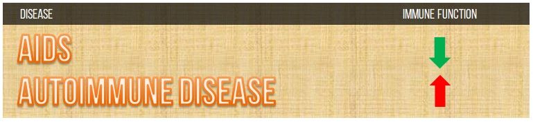
How could the immune system be simultaneously suppressed and in overdrive?
Indeed something is wrong with the theory of autoimmune disease. The first step towards curing a disease is accurately understanding it and what is happening inside the body. Let’s see if we can piece this one together.
Autoimmune Disease More Common in Women
One clue in helping us determine the cause of this disease is that the ratio of women to men who have autoimmune conditions is as high as 10 to 1. Autoimmune disease is up to 10x more prevalent in women than in men. In 1990, UK scientists wrote that “The female to male ratio is 9:1 in systemic lupus erythematosus and 4:1 in rheumatoid arthritis.”[3]
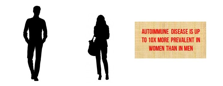
Could estrogen play a role in autoimmune disease?
Estrogen Dominance & “Autoimmune Disease”
It turns out, when you look at some of the most common autoimmune conditions you find that estrogen levels are almost universally increased in patients with these conditions.
- Estrogen levels are significantly increased in patients with rheumatoid arthritis[4]
- Estrogen levels are significantly increased (and testosterone decreased) in patients with lupus[5]
- Estrogen levels are significantly increased in patients with inflammatory bowel disease (IBD)[6]
- Estrogen levels are significantly increased in patients with multiple sclerosis[7]
- Estrogen levels are significantly increased in patients with Hashimoto’s Thyroiditis[8]
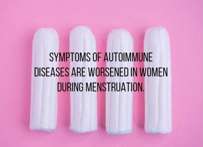
Let’s go over some more evidence of estrogen’s role in this pathology. Symptoms of autoimmune disease are worsened in women during menstruation.[9] Of course, during menstruation in women, estrogen levels are known to be sky high.
Estrogen Treatment Elevates Antibodies
One final proof that estrogen is central to the pathology of autoimmune disease is its relation to autoantibodies. FDA researchers in Bethesda, Maryland showed in a 2001 study that treating mice with estrogen “induces polyclonal B cell activation with increased expression of autoantibodies characteristic of autoimmune diseases.”[10]
Estrogen causes the increased levels of autoantibodies in the blood serum that doctors use to diagnose autoimmune disease.
Why are antibodies increased in the presence of estrogen?
The Truth About Autoantibodies
When a doctor sees “autoantibodies” for a specific tissue on a blood test, he or she thinks that’s evidence the immune system is targeting that tissue. But another hypothesis backed by a mountain of evidence better explains the role of autoantibodies.
Japanese scientists wrote the following in 2009: “Here, we show that B cells, which produce antibodies to damaged tissues, are engaged in the process of wound healing.” In this particular experiment, the researchers used splenectomized mice (mice that don’t have spleens/no B cells/can’t produce autoantibodies) and found that wound healing was delayed. Then the researchers injected B cells into the mice to test the impact on healing. “Transfer of B cells into splenectomized mice restored the wound repair ability.”[11] In other words, antibodies are an essential component for healing damaged tissue; they are the immune system’s clean up crew for removing dead and damaged cellular debris.
“Rather than looking for foreign invading pathogens, this theory says it’s primarily cleaning up messes caused by anything which is pathogenic. In one of the so-called autoimmune brain conditions, people demonstrated that the presence of the antibody to the brain tissue accelerates the recovery. So, cleaning up the mess is really constructive, rather than always being the cause of deterioration.”
– Dr. Ray Peat
Autoantibodies are released to heal damaged tissue caused by estrogen and estrogenic environmental chemicals.
How Does Estrogen Cause Damage?
There are many ways that estrogen damages the body. Let’s go over a few of them now.
Immune System:
Estrogen stimulates antibody production while simultaneously shrinking the thymus gland, which is involved with immunity.[15] But Estrogen also damages the immune system in another way. Estrogen causes chronically elevated free fatty acids in the blood serum, which directly kill white blood cells.[12]
Thyroid:
Estrogen inhibits thyroid function, which plays a major role in today’s common illnesses. American physician Dr. Broda Barnes wrote “If hypothyroid people don’t die young from infectious diseases, such as tuberculosis, they die a little later from cancer or heart disease.”[13]
Adrenals:
Estrogen activates the production of the catabolic stress hormone cortisol.[14] Cortisol is the trigger for the breakdown of body tissues to be used for energy to cope with chemical or environmental stressors. Cortisol’s catabolic effect is one of the primary mechanisms for the damage.seen in autoimmune diseases.
Autoimmune Disease Re-Defined
While the mainstream view of autoimmune disease believes that the antibodies seen in autoimmune patients are indicative of the immune system attacking its own tissues, another hypothesis indicates the pathology is something entirely different.
In “autoimmune disease” environmental chemicals, which exert an estrogenic effect on cells, activate the body’s stress hormones to begin catabolizing its own tissues in order to produce the energy required to deal with the toxic exposure.
A major player in autoimmune disease is cortisol, which destroys the thymus involved in immunity and shuts down the thyroid to slow the metabolism so the body doesn’t eat itself to death. Cortisol is a catabolic hormone that causes tissue breakdown, and it’s this tissue breakdown that leads to a mess of dead cellular debris in the bloodstream which requires cleaning.
In a recent interview Russian researcher Georgi Dinkov summed it up nicely when he said, “Autoantibodies are there to get rid of the dead cellular debris because if it accumulates too much you’ll die of septicemia or some other kind of blood poisoning.”
In summary, the immune system response is occurring in people diagnosed with autoimmune conditions to clear up the debris caused by excessive tissue damage triggered by the stress response. No matter where in the body a person is experiencing symptoms, the disease is always systemic and must be treated as such.
The Smoking Gun: Liver Damage and Autoimmune Disease
And if it’s true that autoimmune disease is caused by exposure to estrogenic environmental chemicals, then one would expect people with autoimmune diseases of all types to almost universally have liver disease. Let’s put the theory to the test. Some have called this ‘The Smoking Gun.’
Liver disease is common in people with virtually all autoimmune conditions:
- Rheumatoid Arthritis[16]
- Lupus[17]
- Inflammatory Bowel Disease (IBD)[18]
- Multiple Sclerosis (MS)[19]
- Type 1 Diabetes[20]
All of these autoimmune conditions are accompanied by high rates of liver damage, which is exactly what you’d expect if these diseases are caused by exposure to large amounts of environmental toxins.
Examples of Environmental Estrogens
The following is a list of some of the most common estrogenic substances within our everyday environment. Reducing your exposure to as many of these factors as possible will reduce your chances of getting autoimmune disease.
- Bisphenol A (BPA)[21]
- Pthalates[22]
- Licorice[23]
- Beer (if it has hops)[24]
- Soy[25]
- Unsaturated vegetable oils[26]
- Iron (food additive)[27]
- Carrageenan (food additive)[28]
- X-rays[29]
- Blue Light[30]
- Dental Amalgams[31]
- Vaccinations
- Mercury is estrogenic[31]
- Aluminum is estrogenic[32]
- Monosodium glutamate is estrogenic[33]
- Pesticides[34]
- Birth control pills[35]
Here is some more information to give you a bigger picture about estrogen’s role in disease and the many other diseases it can cause. In the early 1990s, the Women’s Health Initiative tested supplemental estrogen on women but was forced to stop early because patients began developing dementia, stroke, heart disease and cancer.[36]
Anti-Estrogenic Protective Factors
In addition to avoiding estrogenic chemicals, certain nutrients, drugs and other factors can be used medicinally to counteract the effects of estrogen. The following is a list of anti-estrogenic substances.
Oh and by the way, research has shown that a thymus gland that has been deteriorated by stress can be regenerated using estrogen (aromatase) inhibitors.[44] This study brings hope to anybody who has been damaged by estrogenic substances that their immunity can be restored. Here are some anti-estrogenic substances that can be used to detoxify estrogen from the body.
- Thyroid[37]
- Progesterone[38]
- Red Light Therapy[39]
- Aspirin[40]
- Oranges[41]
- Coconut Oil[42]
- Sodium Bicarbonate[43]
Conclusion
Mainstream medicine has no idea what causes autoimmune disease. They insist the patient’s own immune system has begun attacking their tissues and their chosen remedy for the situation is to administer toxic treatments to try and defeat the naughty immune system. Meanwhile, industry funded studies continue searching for a genetic explanation for the disease, even though a hormonal explanation has already been well established.
Anybody who thinks and examines the evidence for themselves, like we’ve done in this presentation, understands that autoimmune disease is really just a condition of widespread tissue damage caused by chemical activation of the stress response. And to these self-determining people there are no shortages of safe alternative treatments available that work.
The logical way to remedy a situation in which the body is overburdened with estrogenic chemicals is to administer anti-estrogenic medicines that will help the body heal. But instead mainstream medicine uses drugs that are estrogenic themselves and only serve to increase the presence of estrogen. However, nobody is forcing the public to take their remedies.
Effective treatments to counteract estrogen dominance include thyroid hormone, progesterone, Red Light Therapy, Coconut Oil, aspirin and niacinamide. All of these remedies are widely available to anybody who wishes to correct the root cause of the issue.
I hope you learned something from this presentation and that it helps you or someone you love eliminate the pathology that some like to call autoimmune disease.
References
- Malatzky-goshen E, Shoenfeld Y. AIDS and autoimmunity. Autoimmunity. 1989;3(3):201-12.
https://www.ncbi.nlm.nih.gov/pubmed/2491628 - Zandman-goddard G, Shoenfeld Y. HIV and autoimmunity. Autoimmun Rev. 2002;1(6):329-37.
https://www.ncbi.nlm.nih.gov/pubmed/12848988 - Denman AM. Sex hormones, autoimmune diseases, and immune responses. BMJ. 1991;303(6793):2-3.
http://www.ncbi.nlm.nih.gov/pmc/articles/PMC1670230 - Capellino S, Montagna P, Villaggio B, Soldano S, Straub RH, Cutolo M. Hydroxylated estrogen metabolites influence the proliferation of cultured human monocytes: possible role in synovial tissue hyperplasia. Clin Exp Rheumatol. 2008;26(5):903-9.
http://www.ncbi.nlm.nih.gov/pubmed/19032826 - Lahita RG, Bradlow HL, Fishman J, Kunkel HG. Abnormal estrogen and androgen metabolism in the human with systemic lupus erythematosus. Am J Kidney Dis. 1982;2(1 Suppl 1):206-11.
http://www.ncbi.nlm.nih.gov/pubmed/7102669 - Available: https://academic.oup.com/ibdjournal/article/24/2/387/4816930
- Mirmosayyeb O, Badihian S, Manouchehri N, et al. The interplay of multiple sclerosis and menstrual cycle: Which one affects the other one?. Mult Scler Relat Disord. 2018;21:46-50.
https://www.ncbi.nlm.nih.gov/pubmed/29455074 - Arduc A, Aycicek dogan B, Bilmez S, et al. High prevalence of Hashimoto’s thyroiditis in patients with polycystic ovary syndrome: does the imbalance between estradiol and progesterone play a role?. Endocr Res. 2015;40(4):204-10.
https://www.ncbi.nlm.nih.gov/pubmed/25822940 - Available: https://academic.oup.com/ibdjournal/article/24/2/387/4816930
- Verthelyi D. Sex hormones as immunomodulators in health and disease. Int Immunopharmacol. 2001;1(6):983-93.
http://www.ncbi.nlm.nih.gov/pubmed/11407317 - Nishio N, Ito S, Suzuki H, Isobe K. Antibodies to wounded tissue enhance cutaneous wound healing. Immunology. 2009;128(3):369-80.
https://www.ncbi.nlm.nih.gov/pubmed/20067537 - Djurhuus CB, Gravholt CH, Nielsen S, et al. Effects of cortisol on lipolysis and regional interstitial glycerol levels in humans. Am J Physiol Endocrinol Metab. 2002;283(1):E172-7.
https://www.ncbi.nlm.nih.gov/pubmed/12067858 - Mazer NA. Interaction of estrogen therapy and thyroid hormone replacement in postmenopausal women. Thyroid. 2004;14 Suppl 1:S27-34.
https://www.ncbi.nlm.nih.gov/pubmed/15142374 - Woods NF, Mitchell ES, Smith-dijulio K. Cortisol levels during the menopausal transition and early postmenopause: observations from the Seattle Midlife Women’s Health Study. Menopause. 2009;16(4):708-18.
https://www.ncbi.nlm.nih.gov/pmc/articles/PMC2749064 - Norbiato G, Bevilacqua M, Vago T, Clerici M. Glucocorticoids and Th-1, Th-2 type cytokines in rheumatoid arthritis, osteoarthritis, asthma, atopic dermatitis and AIDS. Clin Exp Rheumatol. 1997;15(3):315-23.
https://www.ncbi.nlm.nih.gov/pubmed/9177930 - Mills PR, Macsween RN, Dick WC, More IA, Watkinson G. Liver disease in rheumatoid arthritis. Scott Med J. 1980;25(1):18-22.
https://www.ncbi.nlm.nih.gov/pubmed/7209493 - Kübel D, Tiller M, Mühling T, et al. [Liver disease in systemic Lupus erythematodes – results of an explorative observational study]. Z Gastroenterol. 2018;56(10):1257-1266.
https://www.ncbi.nlm.nih.gov/pubmed/30103221 - Principi M, Iannone A, Losurdo G, et al. Nonalcoholic Fatty Liver Disease in Inflammatory Bowel Disease: Prevalence and Risk Factors. Inflamm Bowel Dis. 2018;24(7):1589-1596.
https://www.ncbi.nlm.nih.gov/pubmed/29688336 - Tremlett H, Seemüller S, Zhao Y, Yoshida EM, Oger JD, Petkau J. Liver test abnormalities in multiple sclerosis: findings from placebo-treated patients. Neurology. 2006;67(7):1291-3.
https://www.ncbi.nlm.nih.gov/pubmed/17030771 - Al-hussaini AA, Sulaiman NM, Alzahrani MD, Alenizi AS, Khan M. Prevalence of hepatopathy in type 1 diabetic children. BMC Pediatr. 2012;12:160.
https://www.ncbi.nlm.nih.gov/pmc/articles/PMC3506494 - Lazúrová Z, Lazúrová I. [The environmental estrogen bisphenol A and its effects on the human organism]. Vnitr Lek. 2013;59(6):466-71.
https://www.ncbi.nlm.nih.gov/pubmed/23808741 - Hong EJ, Ji YK, Choi KC, Manabe N, Jeung EB. Conflict of estrogenic activity by various phthalates between in vitro and in vivo models related to the expression of Calbindin-D9k. J Reprod Dev. 2005;51(2):253-63.
https://www.ncbi.nlm.nih.gov/pubmed/15883486 - Hajirahimkhan A, Simmler C, Yuan Y, et al. Evaluation of estrogenic activity of licorice species in comparison with hops used in botanicals for menopausal symptoms. PLoS One. 2013;8(7).
https://www.ncbi.nlm.nih.gov/pmc/articles/PMC3709979 - Leavy M, Trottmann M, Liedl B, et al. Effects of Elevated β-Estradiol Levels on the Functional Morphology of the Testis – New Insights. Sci Rep. 2017;7:39931.
https://www.ncbi.nlm.nih.gov/pmc/articles/PMC5206739 - Hilakivi-clarke L, Cho E, Clarke R. Maternal genistein exposure mimics the effects of estrogen on mammary gland development in female mouse offspring. Oncol Rep. 1998;5(3):609-16.
https://www.ncbi.nlm.nih.gov/pubmed/9538161 - Reed MJ, Beranek PA, Cheng RW, James VH. Free fatty acids: a possible regulator of the available oestradiol fractions in plasma. J Steroid Biochem. 1986;24(2):657-9.
https://www.ncbi.nlm.nih.gov/pubmed/3702444 - Available at: http://www.ghrnet.org/index.php/ijhr/article/view/1433/1871. Accessed October 3, 2019.
- Misiewicz B, Griebler C, Gomez M, et al. The estrogen antagonist tamoxifen inhibits carrageenan induced inflammation in LEW/N female rats. Life Sci. 1996;58(16):PL281-6.
https://www.ncbi.nlm.nih.gov/pubmed/8614285 - Suman S, Johnson MD, Fornace AJ, Datta K. Exposure to ionizing radiation causes long-term increase in serum estradiol and activation of PI3K-Akt signaling pathway in mouse mammary gland. Int J Radiat Oncol Biol Phys. 2012;84(2):500-7.
https://www.ncbi.nlm.nih.gov/pmc/articles/PMC3580184 - Garcia-saenz A, Sánchez de miguel A, Espinosa A, et al. Evaluating the Association between Artificial Light-at-Night Exposure and Breast and Prostate Cancer Risk in Spain (MCC-Spain Study). Environ Health Perspect. 2018;126(4):047011.
https://www.ncbi.nlm.nih.gov/pubmed/29687979 - Wang YD, Chen XY, Wu YM, Xu D. [Experiment study on the estrogen-like effect of compounds of mercury, chromium and manganese]. Wei Sheng Yan Jiu. 2005;34(1):49-51.
https://www.ncbi.nlm.nih.gov/pubmed/15862022 - Gorgogietas VA, Tsialtas I, Sotiriou N, et al. Potential interference of aluminum chlorohydrate with estrogen receptor signaling in breast cancer cells. J Mol Biochem. 2018;7(1):1-13.
https://www.ncbi.nlm.nih.gov/pmc/articles/PMC6108589 - Zia MS, Qamar K, Hanif R, Khalil M. Effect of monosodium glutamate on the serum estrogen and progesterone levels in female rat and prevention of this effect with diltiazem. J Ayub Med Coll Abbottabad. 2014;26(1):18-20.
https://www.ncbi.nlm.nih.gov/pubmed/25358208 - Mesnage R, Phedonos A, Biserni M, et al. Evaluation of estrogen receptor alpha activation by glyphosate-based herbicide constituents. Food Chem Toxicol. 2017;108(Pt A):30-42.
https://www.ncbi.nlm.nih.gov/pubmed/28711546 - Available: https://www.rxlist.com/birth_control_pills/drugs-condition.htm
- Women’s Health Initiative. Available:
https://www.nhlbi.nih.gov/whi/estro_alone.htm. [May 1, 2019]. - Gregoraszczuk EL, Slomczynska M, Wilk R. Thyroid hormone inhibits aromatase activity in porcine thecal cells cultured alone and in coculture with granulosa cells. Thyroid. 1998;8(12):1157-63.
https://www.ncbi.nlm.nih.gov/pubmed/9920372 - Hardy DB, Janowski BA, Chen CC, Mendelson CR. Progesterone receptor inhibits aromatase and inflammatory response pathways in breast cancer cells via ligand-dependent and ligand-independent mechanisms. Mol Endocrinol. 2008;22(8):1812-24.
https://www.ncbi.nlm.nih.gov/pubmed/18483177 - Höfling DB, Chavantes MC, Juliano AG, et al. Low-level laser in the treatment of patients with hypothyroidism induced by chronic autoimmune thyroiditis: a randomized, placebo-controlled clinical trial. Lasers Med Sci. 2013;28(3):743-53.
https://www.ncbi.nlm.nih.gov/pubmed/22718472 - Yamamoto M, Saito S, Kaise K, Kaise N, Yoshida K, Yoshinaga K. Changes in thyroid hormones by treatment with aspirin and prednisolone in subacute thyroiditis with hyperthyroidism. Tohoku J Exp Med. 1979;127(1):85-95.
https://www.ncbi.nlm.nih.gov/pubmed/760253 - Jeong HJ, Shin YG, Kim IH, Pezzuto JM. Inhibition of aromatase activity by flavonoids. Arch Pharm Res. 1999;22(3):309-12.
https://www.ncbi.nlm.nih.gov/pubmed/10403137 - Scalfi L, Coltorti A, Contaldo F. Postprandial thermogenesis in lean and obese subjects after meals supplemented with medium-chain and long-chain triglycerides. Am J Clin Nutr. 1991;53(5):1130-3.
https://www.ncbi.nlm.nih.gov/pubmed/2021124 - Disthabanchong S, Treeruttanawanich A. Oral sodium bicarbonate improves thyroid function in predialysis chronic kidney disease. Am J Nephrol. 2010;32(6):549-56.
https://www.ncbi.nlm.nih.gov/pubmed/21042013 - Greenstein BD, De bridges EF, Fitzpatrick FT. Aromatase inhibitors regenerate the thymus in aging male rats. Int J Immunopharmacol. 1992;14(4):541-53.
https://www.ncbi.nlm.nih.gov/pubmed/1521922